Prostate biopsy
The most advanced examination in the prostate cancer care
Prostate cancer is the first male cancer and it represents the third cause of death in cancer male patients in France.
Prostate cancer is the most frequent male cancer in developed countries (1,1 million of new cases were identified all over the world in 2012, of which 53000 in France).
Taking care of such a disease is a major public health issue in view of the continuous increase of life expectancy.
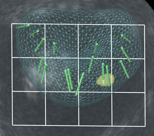
These biopsies are routinely performed with a transrectal approach using ultrasound guidance. Nevertheless this gesture remains relatively blind as ultrasound is more adapted to identify the organ than to spot the tumor which is the most of the time invisible. Twelve samples harmoniously spread within the prostate are usually taken.
The image-guided targeted prostate biopsy
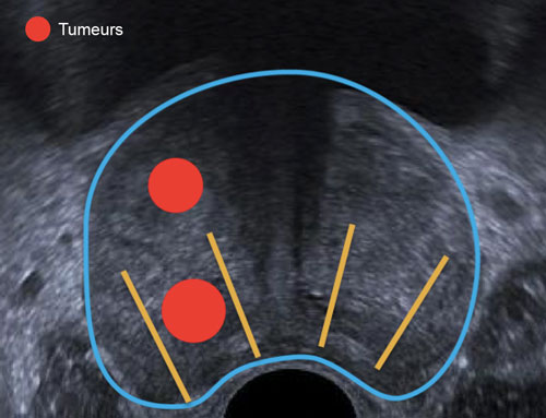
Developments in medical imaging and the advent of multiparametric MRI (Magnetic Resonance Imaging) have made it possible to visualize intra-prostatic tumors who become precise targets for the biopsies.
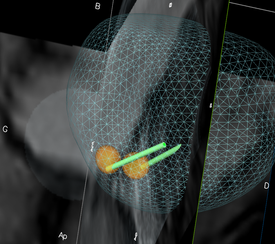
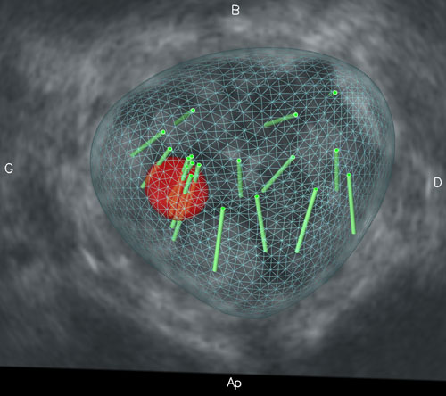
In order to make tumors appear during the ultrasound examination, we are equipped with the Koelis system that uses images fusion to superimpose MRI information with the real time ultrasound image at the moment of the biopsy.
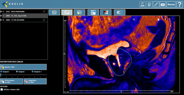
The 3D monitoring system that we use enables a 3 to 4mm accuracy for the biopsy of intra-prostatic targets. It takes into account the movement of the prostate during the process, at the cost of a longer surgery duration.
The current and upcoming advances in prostatic imaging as well as the development of focal treatments are progressively making prostatic biopsies with a 3D transrectal ultrasound and elastic image fusion approach a compulsory step in the initial prostate cancer care.

